When a Brain Mass Is Found By Accident: Patient Gets Life-Saving Brain Surgery

When a rash appeared on his left leg, Tavis Tindall’s first instinct was to ignore it. A former college football player, Tindall still worked out almost daily and followed a healthy diet. “I’ve stayed in tip-top shape since college and never had any health problems,” he says.
However, the rash turned out to be just the first of a series of puzzling issues for which Tindall ended up seeking medical attention. Without what turned out to be an unrelated rash, doctors may not have found two masses in his brain that could have eventually turned life-threatening. One was a brain tumor; the other was a cavernoma, or a small tangle of abnormal blood vessels. Both were located deep within the ventricular system of his brain.
It’s estimated that a little over 87,000 new cases of brain tumors, both cancerous and benign, will be diagnosed in the United States this year. Though Tindall’s tumor fell within a category of slow-growing types called subependymomas, its specific location made it worrisome and potentially dangerous, as did the cavernoma.
“With these types of tumors, we can typically monitor them throughout the patient's life,” says Jennifer Moliterno, MD, Yale Medicine’s chief of neurosurgical oncology for primary brain tumors. “Some patients never experience any trouble from subependymomas,” she says. In those cases, the tumor may never cause symptoms or interfere with the brain’s functions.
But Tindall’s tumor had grown enough to obstruct the ventricles, or the cerebrospinal fluid (CSF)-filled cavities in his brain. The brain has a complex ventricular system, all filled with CSF, which provides a protective cushion around and within the brain, allowing it to float inside the skull. (The ventricles are connected to each other, allowing the CSF to flow between them through a few small openings.)
Tumors such as Tindall’s can interfere with the flow of CSF, essentially stopping it, leading to a potentially life-threatening condition called hydrocephalus. This happens when CSF builds up in the ventricle, leading to excess pressure on and within the brain, and potentially causing brain damage or even death when it occurs acutely, says Dr. Moliterno. (Hydrocephalus is sometimes called “water on the brain.”)
“His lateral ventricle was quite large, but he was not experiencing the typical issues related to hydrocephalus, which led me to believe it had adapted over time to what was likely a slow-growing tumor by expanding in size,” Dr. Moliterno says. “So, although the flow of CSF was blocked, his brain had likely been been able to compensate.” This bought Tindall extra time and likely explained why he wasn’t experiencing symptoms, which can range from persistent headaches to lethargy.
The second mass in Tindall’s brain was another very rare condition called a cerebral cavernous malformation, which affects about 0.2% of the U.S. population. These clusters can develop randomly in individuals, or the condition can be genetic and passed on in families. About one-quarter of people with this condition never experience health problems, but for others the cluster of blood vessels can cause severe symptoms, such as seizures or bleeding in the brain. Bleeding in the ventricle could be particularly worrisome, says Dr. Moliterno.
The path to a brain tumor diagnosis
When Tindall recounts his medical odyssey, he begins with a CrossFit workout in November 2019.
“I was going nonstop from one thing to the next. And at the end of each repetition, I did box jumps,” he says. On his last set, before he jumped onto the three-and-a-half-foot plyometric box, he recalls that a voice in his head said, "Don’t hit your shin."
“What do you think happened? I hit my shin,” Tindall says.
He landed hard on the box’s metal top and his shin began swelling immediately. Tindall finished his workout. “I didn’t ice it or see a doctor or anything,” he says. “I just wanted to let the body heal itself.” Gradually, the swelling went away.
About a month later, the rash appeared. “It was about two inches down and two inches to the right of where I’d struck my leg,” he says. After a few days, his entire left leg turned red and his calf and quadricep muscles began swelling.

"Getting treatment by experienced neurosurgeons who frequently treat rare brain tumor patients can make a difference in care," says Jennifer Moliterno, MD, Yale Medicine’s chief of neurosurgical oncology for primary brain tumors.
Tindall, a lawyer specializing in personal injury, knew enough about medical conditions to guess that his shin injury might have caused deep vein thrombosis; this happens when a blood clot forms in a deep vein. Tindall was aware that sometimes a piece breaks loose, travels through the bloodstream, and has the potential to fatally block blood flow to the lungs.
So, on Christmas Eve, his wife, Kelly, drove him to the emergency department. A doctor found a superficial blood clot in his leg. Not life-threatening, but serious enough that he prescribed blood-thinning medication and told Tindall to take it for a few months.
In February, a few nights before Tindall and Kelly flew to Miami for a long Valentine’s Day weekend, he woke up with a pounding headache. “It felt like my head was about to explode,” he says. He called his hematologist. Since he had no other symptoms, she gave him the go-ahead to fly. “But she told me, ‘When you get back from Florida, we’re going to do an MRI of your brain,’” Tindall recalls.
‘What’s going on?’
On the first night in Miami, Tindall woke up in a panic with a squeezing pressure in his chest. Unsure if he was having a panic attack or a heart attack, he paced in the hotel hallway to calm himself. “My wife found me and was like, ‘What’s going on?’” he says.
They drove to the closest emergency department. A medical team took his vitals, tested his blood, and monitored his heartbeat with an electrocardiogram (EKG) test. Everything came back normal. On a hunch, a physician asked for a computed tomography (CT) scan of his brain.
“That’s when they found the two masses,” Tindall says. The hospital staff assembled a neurosurgery team on the spot. But Tindall chose to fly home for treatment. “I didn’t want to do something impulsively—it’s my brain,” he says.
Back in Southington, Conn., he and his wife asked physician friends to recommend the best neurosurgeon to treat his masses. Two friends—one on the East Coast and the other on the West Coast—simultaneously got back to Tindall and his wife with the same name: Jennifer Moliterno. Tindall called Yale Medicine’s Neurosurgery Department and asked for a consult.
Dr. Moliterno ordered a specialized MRI scan to check the flow of CSF fluid in his brain. She found the subependymoma, which had grown to about the size of a large cherry tomato, was blocking one of the openings in the ventricle about the size of half a pea. “The tumor was about six centimeters deep from his skull and beneath the frontal lobe—and also close to memory fibers,” she says. She told Tindall she would also need to remove the cavernoma as well.
Dr. Moliterno scheduled a surgery for late March of 2020. Then the COVID-19 pandemic hit.
Because Tindall did not have any related symptoms, Dr. Moliterno and he decided to hold off on surgery and instead closely monitor his condition with periodic MRI scans and telehealth visits. Meanwhile, Dr. Moliterno, who continued to operate on emergent and urgent cases during the COVID-19 surge, was prepared to do so with Tindall if any issues developed.
Thankfully, he did not develop any issues, and as the state began to open up for elective surgeries, Dr. Moliterno rescheduled his procedure for mid-June of 2020 (and he safely came off of blood thinners prior to surgery).
Getting proper care for brain tumors
He decided to take charge, to the extent that it was possible. “I am a strong Christian, and I have always enjoyed God’s blessings and protection in my life. Once I made peace with the fact that I needed to have the surgery and that God would protect me during it, I approached it like a sporting event,” Tindall says. He set for himself the goal of walking unassisted the same day as surgery, as well as being able to communicate with his loved ones, friends, and associates that same day.
Before the procedure, Dr. Moliterno met with Tindall to address any lingering questions or concerns, which she considers an important aspect of patient care. “I try my best to understand what my patients are concerned about in an effort to reassure them as I can only imagine how scared they must be,” she says. She told him her “A Team” of neuro-anesthesiologists, nurses, surgical technicians, and residents would be assisting her during the surgery.
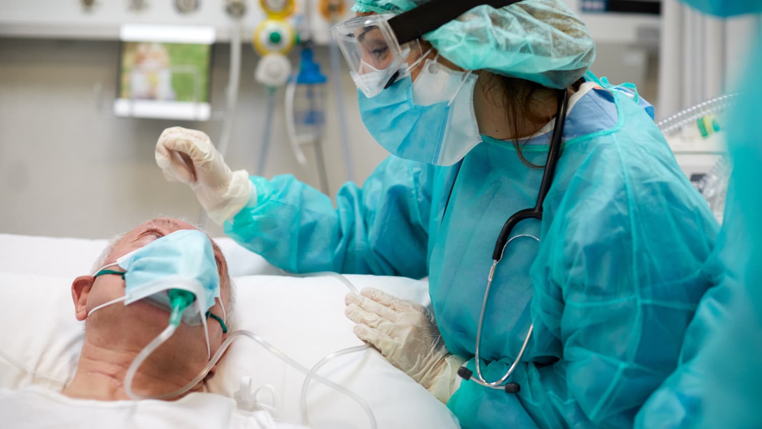
Dr. Moliterno used microsurgical techniques to remove Tindall’s masses. “When working in the ventricle so deep within the brain, it is especially important for a neurosurgeon to be expert with the operative microscope to perform such delicate surgery,” she explains. “Tavis’s surgery is quite complex, and every surgical move matters.”
At the start of the operation, she removed a small part of his skull, a procedure called a craniotomy. Then, Dr. Moliterno used a specialized navigation system to pass a catheter into the ventricle and follow it down to where she encountered the tumor. (Though Yale is the only hospital in the state to have an intraoperative magnetic resonance imaging [iMRI] machine, she did not need that technology for this surgery.)
Prior to the procedure, she’d estimated that Tindall’s surgery could last six hours, but she was able to finish in two. “I do this a lot, so I know how to move quickly but safely,” Dr. Moliterno says.

“I didn’t want to do something impulsively—it’s my brain,” says Tavis Tindall, on his search for the right neurosurgeon.
Getting treatment by experienced neurosurgeons who frequently treat rare brain tumor patients can make a difference in care, Dr. Moliterno says. “A center like ours—with a high volume of brain tumor surgeries, along with specialized, dedicated brain tumor neurosurgeons—really matters in terms of how patients do,” she says. “We serve as a destination center for patients with more complex brain tumors in the region and beyond.”
For his part, Tindall is thankful to God for the freak sports injury, which led to the discovery of the tumors, and for leading him to such a skilled and caring surgeon as Dr. Moliterno and the neurosurgery department team. “Dr. Moliterno and everyone on her team were phenomenal—the whole experience was great,” he says.
Tindall recovered quickly from the surgery. He met his goal of walking without help the same day, as well as communicating with friends and loved ones the night after the surgery. Just five days later, he walked a couple miles to raise money for a charity that provides meals to military veterans, poor families, and the homeless in the greater Waterbury area. Following the surgery, Tindall says he has experienced no memory loss issues, no impact upon his thinking skills, and “no other negative effects of any sort upon his brain.” And only eight days after the surgery—on his 49th birthday—Tindall was able to conduct a civil pre-trial on one of his client’s cases.
Tindall recalls that just before the surgery, he found an old CT scan of his brain taken in 2013.
The two masses weren’t there. That meant the masses, which are usually slow-growing, had grown relatively quickly.
“That was another sign to me that surgery was the right choice,” Tindall says. “I recently told Dr. Moliterno that I literally thank God every day in prayer for Him helping me discover these tumors and putting Dr. Moliterno in my life to perform this crucial surgery. Her incomparable skills as a surgeon are matched only by her caring heart and tremendous bedside manner.”