Biopsy
Definition
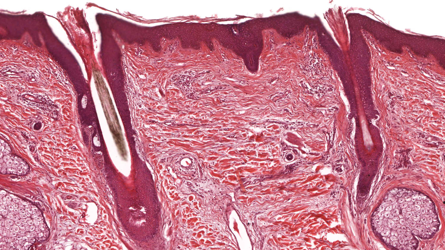
A biopsy is a medical procedure in which a small sample of tissue is removed from a specific area of the body for examination under a microscope. This diagnostic technique helps doctors determine the presence, cause, or extent of a disease, such as cancer or infection.
Related Fact Sheets
Showing 6 of 21
Related News
Related Departments
Showing 3 of 11
Anatomic Pathology
Whereas laboratory scientists typically analyze blood or other fluids from patients, anatomic pathologists evaluate tissue specimens, which include cervical scrapings (i.e., PAP smears), biopsies, surgical resections, and whole-body evaluations at autopsy. Anatomic Pathology has three major divisions: Surgical Pathology. This is the examination of tissue removed as a biopsy or as part of a surgical procedure. When a biopsy is done (typically a small sampling of a lesion by incision or by an invasive technique like a core needle biopsy or endoscopy), the pathologist determines what disease process is present, and/or the extent of disease. In all cases, pathologists make their diagnoses by examining thin slices of the lesion under a microscope. Pathologists often also use a variety of specialized molecular techniques to further refine the diagnosis and to predict how the disease may respond to various types of treatment. In this way, the pathologist helps guide any subsequent therapy the patient may need. Cytopathology: This is the examination of very small amounts of tissue removed by scraping a surface, or by aspiration through a fine needle. Obtaining a cytopathology specimen is typically less invasive than obtaining a surgical pathology specimen, so these procedures can be performed in a clinic or a physician’s office. Cytopathologists examine individual cells and small collections of cells to assess for the presence or absence of malignancy. Autopsy Pathology: This is the anatomic examination of a deceased patient to determine what diseases were present and how extensive they were, and to assemble these findings into an explanation for why the patient died. Autopsy examination can answer questions family members may have about the patient’s death, but can also increase understanding of disease for the physicians caring for the patient.Breast Pathology
The Breast Pathology program specializes in interpreting and evaluating core needle breast biopsies, incisional and excisional biopsies, lumpectomy or partial and total mastectomy specimens, sentinel lymph nodes, axillary dissections, prophylactic and oncoplastic reduction mammoplasty, and neoadjuvant breast cases. The faculty is board-certified in anatomic pathology and many of the pathologists also have subspecialty expertise in breast pathology. We provide state-of-the-art diagnostic services focused on precise diagnoses of both benign and malignant breast diseases. In cases with malignant diagnosis, additional ancillary studies for receptors are reported with two- to three-day turnaround times. We work closely with our clinical team to ensure that our patients get accurate and reliable results that are critical to patient care. The first question a breast pathologist seeks to answer when reading a breast biopsy is whether cancer is present. But the information included in the pathology report goes far beyond the “yes” or “no” diagnosis. Even if the biopsy is benign, we need to ensure that the calcifications seen on imaging correlate with calcifications seen on the core biopsy pathology specimen. The earliest stage of breast cancer, called ductal carcinoma in situ (DCIS), is usually detected in this manner. Discordance between pathology and imaging is addressed by means of communication between the radiologist and pathologist in a radiology-pathology conference or by individual communication. A pathology report always has a detailed visual morphologic description of the tumor. The initial biopsy report includes information on whether the cancer is confined within the ducts (in situ carcinoma) or has breached the duct wall and invaded into the adjacent stroma (invasive carcinoma). We provide intraoperative consultation on sentinel lymph nodes in the frozen section suite to detect metastases, which will then help the surgeon in planning patient management. Additional details will be studied and added to the pathological report after mastectomy and sentinel node biopsy to determine whether the cancer has metastasized or spread to any lymph nodes. Tumor size, histologic grade, and lymph node status are prognostic indicators that provide valuable information about the likely clinical outcome. For example, a patient whose tumor is well-differentiated and has negative margins, i.e., has clear margins, has a better prognosis than a patient whose tumor is one that is poorly differentiated and is present at the margin.Cutaneous Oncology Program
We offer sophisticated approaches for the management of skin cancers, ranging from the most common to the most complicated or rare. At the heart of our program is microscopically controlled excision, known asMohs surgery, the most successful treatment to cure recurrent or other high-risk skin cancers. We have been offering the procedure since 1998, and today, our physicians perform 3,500 Mohs surgeries each year. Mohs surgery has revolutionized the treatment of such skin cancers as basal cell carcinoma and squamous cell carcinoma, and the chances of lasting, disfiguring scars are minimized. In this procedure, the cancer is removed layer by layer, and, after each step, the tissue is examined under a microscope, allowing our dermatologists to confirm that all of the cancer cells have been eliminated as the surgery progresses. This maximizes the chances of removing all of the abnormal cells while still preserving as much of the normal skin tissue as possible.