Corneal Abrasion
Overview
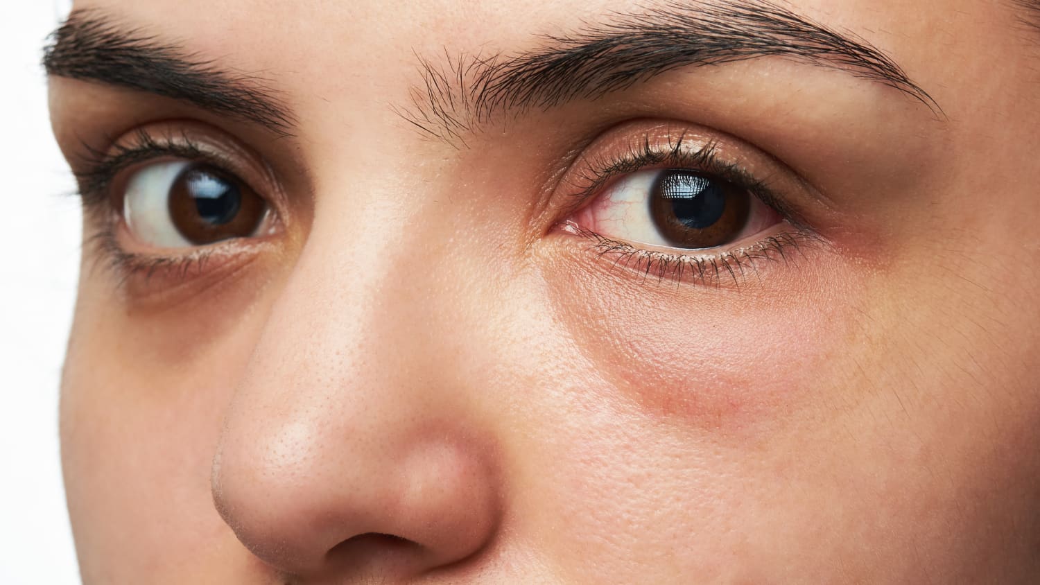
It feels like a grain of sand is trapped in your eye that won't come out, and your eye is teary, stinging when you look into the light. If you have these symptoms, you could have a corneal abrasion, the most common trauma to the eye. A corneal abrasion affects the cornea, the outermost layer of the eye, which is vulnerable to scratches and pokes that can tear this delicate tissue.
It is important that abrasions are treated properly, says Jessica H. Chow, MD, a physician with Yale Medicine Ophthalmology. We have specialists with training in treating the cornea, state-of-the-art diagnostic tools and, if it becomes necessary, access to the latest, most advanced eye treatments.
What are the symptoms of corneal abrasion?
Corneal abrasions can cause significant pain. “The cornea has among the highest densities of nerve fibers in the human body, so even a very small abrasion can be very painful,” says Dr. Chow.
Redness, tearing, sensitivity to light and the sensation that something is in the eye are also signs and symptoms of corneal abrasion.
What are the risk factors for corneal abrasion?
Everyone is at risk for suffering an abrasion with direct trauma to the eye. However, certain people may have an eye condition that predisposes them to recurrent corneal erosions.
How can corneal abrasions be prevented?
By keeping fingers away from the eyes, a person can reduce the risk of accidentally scratching the cornea. If an object gets in the eye, rinse the eye with clean water or saline solution, but do not rub the eye. If a foreign object is visible on the cornea, do not try to remove it, and call your doctor instead.
How are corneal abrasions diagnosed?
Corneal abrasions are usually not visible to the naked eye, but an ophthalmologist can clearly see an abrasion with a slit-lamp biomicroscope, a tool that is commonly used during a routine eye exam. The slit-lamp microscope combines a high-powered light source with a microscope, so it allows the ophthalmologist to examine the eye closely.
Corneal abrasions are also diagnosed using a special dye and light, especially in emergency departments or health care offices without access to slit-lamp biomicroscopes. After drops of fluorescein dye are inserted in the eye, the doctor shines a blue light on the eye. Any abrasions on the eye will appear green.
How are corneal abrasions treated?
In people who are in good general health, most typical corneal abrasions can heal on their own within 24 to 48 hours. A doctor may prescribe antibiotic eye drops or ointment. Because the cornea is so sensitive, simply opening and closing the eye over the abrasion may be painful.
“Keeping the eye closed as much as possible in the first day or two after the injury can help with the pain,” says Dr. Chow. In some cases, the ophthalmologist will put an antibiotic or anti-inflammatory ointment into the eye and then use a patch to keep the eye closed.
What makes Yale Medicine’s approach to treating corneal abrasions unique?
Although corneal abrasions are very common and usually heal on their own, the access to state-of-the-art diagnostic and therapeutic techniques provided by Yale Medicine should bolster patients’ confidence that they are receiving the best available medical care.